Ultrasound Machine
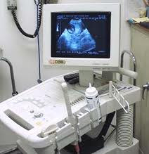
Ultrasound machines represent a non-invasive method for obtaining crucial
medical information, surpassing traditional diagnostic tools and techniques.
These machines, equipped with probes, are employed either internally or
externally to capture clear images of the fetus and its development within
the womb.
The applications of ultrasound machines are diverse, frequently utilized in
obstetrics, echocardiography, biopsy, and various other medical procedures,
enhancing the safety and precision of medical practice. Diagnostic
sonography, also known as ultrasonography, employs ultrasound waves to
visualize subcutaneous body structures like tendons, muscles, joints,
vessels, and internal organs, aiding in the identification of potential
pathology or lesions.
Obstetric sonography, a widely recognized application of ultrasound, is
commonly employed during pregnancy. In physics, "ultrasound" refers to sound
waves with frequencies above the audible range of human hearing,
approximately 20 kHz. Diagnostic ultrasound typically operates within the
frequency range of 2 to 18 megahertz, with experimental frequencies reaching
up to 50–100 megahertz in specialized techniques like biomicroscopy. The
choice of frequency involves a balance between spatial resolution and
imaging depth: lower frequencies offer deeper imaging but lower resolution,
while higher frequencies provide better resolution but shallower penetration
into the body.
Sonography finds extensive use in medicine, enabling both diagnostic and
therapeutic procedures. Sonographers, trained medical professionals, perform
scans using handheld probes known as transducers. These scans are then
interpreted by Radiologists or Cardiologists, specialists in medical imaging
or cardiac ultrasonography, respectively. Sonography effectively images soft
tissues such as muscles, tendons, testes, breast tissue, and the neonatal
brain at higher frequencies (7–18 MHz), offering superior resolution. Deeper
structures like the liver and kidneys are imaged at lower frequencies (1–6
MHz), providing greater penetration but lower resolution.
undo Medical Equipment